YUHINA
  • Home
  • Process
  • Outputs
  • Pricing
English▾
  • English
  • العربية
  • Français
  • Español
  • Deutsch
  • Português
  • Italiano
  • हिन्दी
  • 中文
  • 日本語
  • 한국어
  • Türkçe
  • Русский
  • Bahasa Indonesia
Sign in

How can we help?

Pick what it's about. The answer may already be below.

Something in the app isn't working the way it should.

My verification code hasn't arrived.+

YUHINA sends a 6-digit code to the address you signed up with. Check your spam or promotions folder. If it still isn't there after a few minutes, email support from that address.

Why can't I generate anything?+

You need a verified email first. A new account includes one free chapter with every output, once. After that, generating spends your plan's monthly usage or pay-as-you-go credit, so you need a plan or some credit to keep going.

My PDF gave weak results.+

Each PDF is one chapter, up to 60 pages. A digital PDF with text you can select gives the clearest lessons; blurry, dark, skewed or handwritten scans give weaker ones.

How do I delete my account or my files?+

The app's privacy notice explains how to export or permanently delete your account and everything in it. If something doesn't work, email support.

Still stuck? Email support, from the address on your account:

support@yuhina.ai

Buying for a school or a company? Talk to our enterprise team: enterprise@yuhina.ai

Sample library · Anatomy and Physiology

Heart Anatomy: video lessons, quiz and flashcards from one textbook chapter

What YUHINA made from one anatomy and physiology chapter: 4 narrated video lessons with slides, 110 questions, 32 flashcards, a mind map and a podcast.

  • 4 lessons
  • 60 slides
  • 110 questions
  • 32 flashcards
  • 43-minute podcast

Generated by YUHINA on 2026-11-10 from a 16-page PDF and shown unedited.

Source: section 19.1 of chapter 19, “The Cardiovascular System: The Heart”, from Anatomy and Physiology (1st edition) by J. Gordon Betts, Kelly A. Young et al., OpenStax, licensed CC BY 4.0. Anatomy and Physiology. Access the book for free at openstax.org. Changes: YUHINA turned the text into lessons, slides, narration, questions, flashcards, a mind map and a podcast.

Try it with your own chapterSee pricing

The narrated video

The first 60 seconds of lesson 1. The chapter came out as 4 video lessons, about 42 minutes in all.

The first slides of lesson 1

  • Slide 1 of lesson 1
  • Slide 2 of lesson 1
  • Slide 3 of lesson 1

The lessons

YUHINA split the chapter into lessons. Each one has its own summary and quiz, shown below as generated.

Lesson 1: Heart Location, Position, and Surface Anatomy

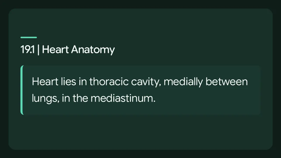
The human heart lies within the thoracic cavity, medially between the lungs, in a space known as the mediastinum.

A tough membrane called the pericardium, or pericardial sac, separates the heart from other mediastinal structures, and the heart sits in its own space called the pericardial cavity.

The dorsal surface of the heart lies near the bodies of the vertebrae, while its anterior surface sits deep to the sternum and costal cartilages.

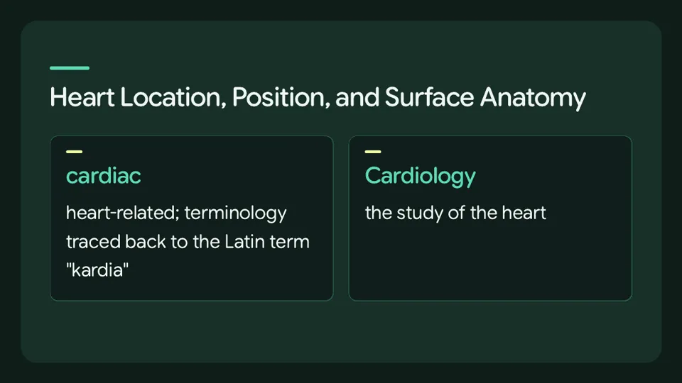
cardiac: heart-related; terminology traced back to the Latin term "kardia"

Cardiology: the study of the heart

base: the superior surface of the heart, to which the superior and inferior venae cavae, aorta and pulmonary trunk are attached

apex: the inferior tip of the heart, lying just to the left of the sternum between the junction of the fourth and fifth ribs near their articulation with the costal cartilages

The right side of the heart is deflected anteriorly, and the left side is deflected posteriorly.

cardiac notch: a depression in the medial surface of the inferior lobe of the left lung reflecting the slight deviation of the apex to the left

19.1 | Heart Anatomy — selected points

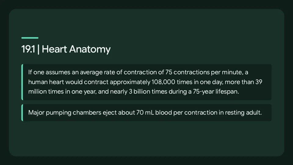
Assuming an average rate of contraction of 75 contractions per minute, a human heart would contract approximately 108,000 times in one day, more than 39 million times in one year, and nearly 3 billion times during a 75-year lifespan.

In a resting adult, each of the major pumping chambers of the heart ejects approximately 70 mL of blood per contraction.

Location of the Heart

The great veins, namely the superior and inferior venae cavae, and the great arteries, namely the aorta and pulmonary trunk, are attached to the superior surface of the heart, which is called the base.

Within the mediastinum, the heart is separated from the other mediastinal structures by a tough membrane known as the pericardium, or pericardial sac, and sits in its own space called the pericardial cavity.

The inferior tip of the heart, the apex, lies just to the left of the sternum between the junction of the fourth and fifth ribs near their articulation with the costal cartilages.

The slight deviation of the apex to the left is reflected in a depression in the medial surface of the inferior lobe of the left lung, called the cardiac notch.

CPR

Applying pressure with the flat portion of one hand on the sternum in the area between the line at T4 and T9 makes it possible to manually compress the blood within the heart enough to push some of that blood into the pulmonary and systemic circuits.

Current standards call for compressing the chest at least 5 cm deep and at a rate of 100 compressions per minute, a rate equal to the beat in "Staying Alive," recorded in 1977 by the Bee Gees.

At this stage, the emphasis is on performing high-quality chest compressions rather than providing artificial respiration, and CPR is generally performed until the patient regains spontaneous contraction or is declared dead by an experienced healthcare professional.

When performed by untrained or overzealous individuals, CPR can result in broken ribs or a broken sternum and can inflict additional severe damage on the patient, and if the hands are placed too low on the sternum it is also possible to manually drive the xiphoid process into the liver, a consequence that may prove fatal for the patient.

Shape and Size of the Heart — selected points

The shape of the heart is similar to a pinecone, being rather broad at the superior surface and tapering to the apex.

A typical heart is approximately the size of a fist, measuring 12 cm (5 in) in length, 8 cm (3.5 in) in width and 6 cm (2.5 in) in thickness.

Given the size difference between most members of the sexes, a female heart weighs approximately 250–300 grams (9 to 11 ounces) and a male heart approximately 300–350 grams (11 to 12 ounces).

Hearts of athletes can pump blood more effectively at lower rates than those of nonathletes.

Enlarged hearts are not always a result of exercise; they can result from pathologies, such as hypertrophic cardiomyopathy.

The cause of an abnormally enlarged heart muscle is unknown, but the condition is often undiagnosed and can cause sudden death in apparently otherwise healthy young people.

Chambers and Circulation through the Heart

Each of the upper chambers, the right atrium (plural = atria) and the left atrium, acts as a receiving chamber and contracts to push blood into the lower chambers, the right ventricle and the left ventricle.

Highly oxygenated blood returning from the pulmonary capillaries in the lungs passes through a series of vessels that join together to form the pulmonary veins, the only post-natal veins in the body that carry highly oxygenated blood.

These vessels in turn branch many times before reaching the pulmonary capillaries, where gas exchange occurs: Carbon dioxide exits the blood and oxygen enters.

Eventually, these vessels will lead to the systemic capillaries, where exchange with the tissue fluid and cells of the body occurs.

In this case, oxygen and nutrients exit the systemic capillaries to be used by the cells in their metabolic processes, and carbon dioxide and waste products will enter the blood.

The blood exiting the systemic capillaries is lower in oxygen concentration than when it entered.

Quiz (7 questions)

  1. Which term matches this description: “the study of the heart”?

    • Cardiology
    • Apex
    • Base
    • Cardiac
    Show the answer

    Cardiology. Cardiology is the study of the heart, and cardiologists are the physicians who deal primarily with the heart. The apex is the inferior tip of the heart, an anatomical landmark rather than a field of study. The base is the superior surface of the heart where the great vessels attach, not a discipline. Cardiac is an adjective meaning heart-related, not the study of the heart.

  2. Which term is defined as “heart-related; terminology traced back to the Latin term "kardia"”?

    • Hypertrophy
    • Cardiac
    • Pericardium
    • Cardiology
    Show the answer

    Cardiac. Cardiac means heart-related, and this terminology can be traced back to the Latin term kardia. Hypertrophy is the enlargement of individual cells through added protein myofilaments, not a term for heart-related. Cardiology is the study of the heart, a field rather than the adjective itself. The pericardium is the tough membrane that surrounds the heart, not the heart-related term derived from kardia.

  3. Which term is defined as “the superior surface of the heart, to which the superior and inferior venae cavae, aorta and pulmonary trunk are attached”?

    • Cardiac
    • Apex
    • Base
    • Cardiology
    Show the answer

    Base. The base is the superior surface of the heart, located at the level of the third costal cartilage, where the superior and inferior venae cavae, the aorta and the pulmonary trunk attach. The apex is the inferior tip of the heart near the fourth and fifth ribs, not the superior surface. Cardiac is an adjective meaning heart-related, not a surface of the heart. Cardiology is the study of the heart, not an anatomical region.

  4. True or false: The pulmonary veins conduct blood into the left atrium, which pumps it into the left ventricle, which pumps oxygenated blood into the pulmonary trunk and the branches of the pulmonary circuit.

    • True
    • False
    Show the answer

    False. The statement is false because the left ventricle pumps oxygenated blood into the aorta and on to the branches of the systemic circuit, not into the pulmonary trunk. The pulmonary trunk receives deoxygenated blood from the right ventricle and leads to the lungs. The pulmonary veins do carry oxygenated blood into the left atrium, and the left atrium does pump it into the left ventricle, but the destination of the left ventricle's output is the systemic circulation.

  5. True or false: The right atrium and left atrium act as receiving chambers and contract to push blood into the lower chambers, the right ventricle and left ventricle.

    • True
    • False
    Show the answer

    True. The statement is true: the right and left atria are the upper receiving chambers, and they contract to push blood into the lower chambers, the right and left ventricles. The ventricles are the primary pumping chambers that propel blood to the lungs or the rest of the body. This division of labor lets the atria fill and then deliver blood to the ventricles for ejection.

  6. True or false: The pulmonary circuit transports blood to and from the lungs, where it picks up oxygen and delivers carbon dioxide for exhalation.

    • True
    • False
    Show the answer

    True. The statement is true: the pulmonary circuit transports blood to and from the lungs, where it picks up oxygen and delivers carbon dioxide for exhalation. The systemic circuit is the separate route that carries oxygenated blood to body tissues and returns deoxygenated blood and carbon dioxide to the heart. Together the two circuits ensure oxygen delivery and waste removal throughout the body.

  7. Which statement defines “apex”?

    • the superior surface of the heart, to which the superior and inferior venae cavae, aorta and pulmonary trunk are attached
    • a depression in the medial surface of the inferior lobe of the left lung reflecting the slight deviation of the apex to the left
    • addition of protein myofilaments that increase the size of individual cells without increasing their numbers
    • the inferior tip of the heart, lying just to the left of the sternum between the junction of the fourth and fifth ribs near their articulation with the costal cartilages
    Show the answer

    the inferior tip of the heart, lying just to the left of the sternum between the junction of the fourth and fifth ribs near their articulation with the costal cartilages. The apex is the inferior tip of the heart, lying just to the left of the sternum between the junction of the fourth and fifth ribs near their articulation with the costal cartilages. The superior surface where the venae cavae, aorta and pulmonary trunk attach is the base, not the apex. The depression in the medial surface of the inferior lobe of the left lung is the cardiac notch, not the apex. The addition of protein myofilaments that enlarges cells without increasing their number is hypertrophy, not the apex.

Lesson 2: Pulmonary and Systemic Circulation and Heart Chambers

A septum is a wall or partition that divides the heart into chambers.

The interatrial septum lies between the two atria, and the interventricular septum lies between the two ventricles.

The interatrial septum bears an oval-shaped depression called the fossa ovalis, which is a remnant of an opening in the fetal heart known as the foramen ovale.

The septum between the atria and ventricles is the atrioventricular septum.

A valve is a specialized structure that ensures one-way flow of blood; the valves between the atria and ventricles are known generically as atrioventricular valves, and the valves at the openings leading to the pulmonary trunk and aorta are known generically as semilunar valves.

Because these openings and valves structurally weaken the atrioventricular septum, the remaining tissue is heavily reinforced with dense connective tissue called the cardiac skeleton, or skeleton of the heart.

Membranes — selected points

The membrane that directly surrounds the heart and defines the pericardial cavity is called the pericardium or pericardial sac, and the pericardium consists of two distinct sublayers: the sturdy outer fibrous pericardium and the inner serous pericardium.

The fibrous pericardium is made of tough, dense connective tissue that protects the heart and maintains its position in the thorax.

In most organs within the body, visceral serous membranes such as the epicardium are microscopic, but in the case of the heart the layer is not microscopic but rather macroscopic, consisting of a simple squamous epithelium called a mesothelium, reinforced with loose, irregular, or areolar connective tissue that attaches to the pericardium.

The pericardial cavity, filled with lubricating serous fluid, lies between the epicardium and the pericardium.

Heart: Cardiac Tamponade — selected points

If excess fluid builds within the pericardial space, it can lead to a condition called cardiac tamponade, or pericardial tamponade.

With each contraction of the heart, more fluid—in most instances, blood—accumulates within the pericardial cavity.

In order to fill with blood for the next contraction, the heart must relax, but the excess fluid in the pericardial cavity puts pressure on the heart and prevents full relaxation, so the chambers within the heart contain slightly less blood as they begin each heart cycle.

Over time, less and less blood is ejected from the heart.

If the fluid builds up slowly, as in hypothyroidism, the pericardial cavity may be able to expand gradually to accommodate this extra volume, and some cases of fluid in excess of one liter within the pericardial cavity have been reported.

Rapid accumulation of as little as 100 mL of fluid following trauma may trigger cardiac tamponade.

Other common causes include myocardial rupture, pericarditis, cancer, or even cardiac surgery, and premature removal of drainage tubes, for example following cardiac surgery, or clot formation within these tubes are causes of this condition.

Untreated, cardiac tamponade can lead to death.

Surface Features of the Heart — selected points

Auricles are relatively thin-walled structures that can fill with blood and empty into the atria or upper chambers of the heart, and they may also be referred to as atrial appendages.

A series of fat-filled grooves, each known as a sulcus (plural sulci), is also prominent along the superior surfaces of the heart, and major coronary blood vessels are located in these sulci.

The deep coronary sulcus is located between the atria and ventricles.

Located between the left and right ventricles are two additional sulci that are not as deep as the coronary sulcus.

The anterior interventricular sulcus is visible on the anterior surface of the heart, whereas the posterior interventricular sulcus is visible on the posterior surface of the heart.

Layers

The wall of the heart is composed of three layers of unequal thickness.

From superficial to deep, these layers are the epicardium, the myocardium, and the endocardium.

Septa of the Heart — selected points

The term septum comes from Latin meaning “something that encloses,” and in the heart a septum (plural septa) is a wall or partition that divides the heart into chambers.

Septa are physical extensions of the myocardium and are lined with endocardium.

The interatrial septum is located between the two atria.

Normally in an adult heart, the interatrial septum bears an oval-shaped depression called the fossa ovalis, which is a remnant of a fetal opening called the foramen ovale.

The foramen ovale allowed blood in the fetal heart to pass directly from the right atrium to the left atrium, allowing some blood to bypass the pulmonary circuit.

Within seconds after birth, a flap of tissue called the septum primum, which previously acted as a valve, closes the foramen ovale and establishes the typical cardiac circulation pattern.

A second septum, the interventricular septum, lies between the two ventricles; unlike the interatrial septum, it is normally intact after its formation during fetal development, and it is substantially thicker than the interatrial septum because the ventricles generate far greater pressure when they contract.

The valves between the atria and ventricles are known generically as atrioventricular valves, while the valves at the openings that lead to the pulmonary trunk and aorta are known generically as semilunar valves.

The cardiac skeleton also provides an important boundary in the heart electrical conduction system.

The septum between the atria and ventricles is known as the atrioventricular septum.

It is marked by the presence of four openings that allow blood to move from the atria into the ventricles and from the ventricles into the pulmonary trunk and aorta.

Located in each of these openings between the atria and ventricles is a valve, a specialized structure that ensures one-way flow of blood.

Quiz (6 questions)

  1. “The innermost layer of the heart wall, joined to the myocardium with a thin layer of connective tissue.” — which term does this describe?

    • Foramen ovale
    • Atrioventricular septum
    • Valve
    • Endocardium
    Show the answer

    Endocardium. The endocardium is the innermost layer of the heart wall, joined to the myocardium by a thin layer of connective tissue. The foramen ovale is a fetal opening between the atria, not a layer of the heart wall. The atrioventricular septum is the partition between the atria and ventricles, not a lining layer. A valve is a structure that ensures one-way flow, not the innermost layer.

  2. Which term matches this description: “An opening in the fetal heart, of which the fossa ovalis is a remnant.”?

    • Foramen ovale
    • Septum
    • Sulcus (plural = sulci)
    • Fossa ovalis
    Show the answer

    Foramen ovale. The foramen ovale is the opening in the fetal heart that allowed blood to pass from the right atrium to the left atrium, and the fossa ovalis is its remnant. A sulcus is a fat-filled groove on the heart's surface containing coronary vessels, not an opening. A septum is a wall or partition dividing the heart into chambers, not an opening. The fossa ovalis is the remnant depression, not the original opening itself.

  3. “A specialized structure that ensures one-way flow of blood.” — which term does this describe?

    • Endocardium
    • Valve
    • Foramen ovale
    • Fossa ovalis
    Show the answer

    Valve. A valve is a specialized structure that ensures one-way flow of blood through the heart. The endocardium is the innermost lining of the heart wall, not a one-way flow structure. The foramen ovale is a fetal opening between the atria, not a valve. The fossa ovalis is a remnant depression on the interatrial septum, not a valve.

  4. True or false: Major coronary blood vessels are located in the sulci.

    • True
    • False
    Show the answer

    True. True. The sulci are fat-filled grooves on the heart's surface, and major coronary blood vessels are located in these grooves. The deep coronary sulcus lies between the atria and ventricles, and the anterior and posterior interventricular sulci are visible on the anterior and posterior surfaces, respectively.

  5. True or false: Contraction of the myocardium pumps blood through the heart and into the major arteries.

    • True
    • False
    Show the answer

    True. True. The myocardium is the middle and thickest layer of the heart wall, made largely of cardiac muscle cells, and its contraction pumps blood through the heart and into the major arteries. The epicardium is the outermost layer, and the endocardium is the innermost lining, but neither is responsible for pumping blood.

  6. True or false: The endothelium may regulate the growth patterns of cardiac muscle cells throughout life.

    • True
    • False
    Show the answer

    True. True. The endothelium, the simple squamous epithelium lining the endocardium and coronary capillaries, may regulate the growth patterns of cardiac muscle cells throughout life. It also plays roles in regulating contraction and ionic concentrations in surrounding tissue fluids.

Lesson 3: Congenital Heart Defects

A patent foramen ovale is a very common form of interatrial septum pathology in which the septum primum does not close at birth and the fossa ovalis is unable to fuse.

A patent foramen ovale is normally detected by auscultation of a heart murmur, an abnormal heart sound, and confirmed by imaging with an echocardiogram.

In nonlife-threatening cases, monitoring the condition is better than risking heart surgery to repair and seal the opening.

Coarctation of the aorta is a congenital abnormal narrowing of the aorta normally located at the insertion of the ligamentum arteriosum, the remnant of the fetal shunt called the ductus arteriosus.

A patent ductus arteriosus is a congenital condition in which the ductus arteriosus fails to close.

Septal defects are not uncommon in individuals and may be congenital or caused by various disease processes, and cyanosis is a blue or purple coloration of the skin, especially when active, caused by an insufficient supply of oxygenated blood.

The right atrium serves as the receiving chamber for blood returning to the heart from the systemic circulation.

The right auricle also has pectinate muscles. The left atrium does not have pectinate muscles except in the auricle.

Heart: Heart Defects — selected points

As much as 20–25 percent of the general population may have a patent foramen ovale, but fortunately most have the benign, asymptomatic version.

Despite its prevalence in the general population, the causes of patent ovale are unknown and there are no known risk factors.

A patent ductus arteriosus is a congenital condition in which the ductus arteriosus fails to close, and failure of the ductus arteriosus to close results in blood flowing from the higher pressure aorta into the lower pressure pulmonary trunk.

Symptoms of a patent ductus arteriosus include shortness of breath (dyspnea), tachycardia, enlarged heart, a widened pulse pressure, and poor weight gain in infants, and if untreated the condition can result in congestive heart failure.

Tetralogy of Fallot is a congenital condition that may also occur from exposure to unknown environmental factors and occurs when there is an opening in the interventricular septum caused by blockage of the pulmonary trunk, normally at the pulmonary semilunar valve.

Symptoms of tetralogy of Fallot include a distinct heart murmur, low blood oxygen percent saturation, dyspnea or difficulty in breathing, polycythemia, broadening (clubbing) of the fingers and toes, and in children, difficulty in feeding or failure to grow and develop, and tetralogy of Fallot is the most common cause of cyanosis following birth.

Septal defects are commonly first detected through auscultation, listening to the chest using a stethoscope, and this is often followed by medical imaging to confirm or rule out a diagnosis, while in many cases treatment may not be needed.

The term “tetralogy” is derived from the four components of the condition, although only three may be present in an individual patient: pulmonary infundibular stenosis (rigidity of the pulmonary valve), overriding aorta (the aorta is shifted above both ventricles), ventricular septal defect (opening), and right ventricular hypertrophy (enlargement of the right ventricle).

Tetralogy of Fallot occurs in approximately 400 out of one million live births.

Normal treatment involves extensive surgical repair, including the use of stents to redirect blood flow and replacement of valves and patches to repair the septal defect, but the condition has a relatively high mortality.

Survival rates are currently 75 percent during the first year of life; 60 percent by 4 years of age; 30 percent by 10 years; and 5 percent by 40 years.

In the case of severe septal defects, including both tetralogy of Fallot and patent foramen ovale, failure of the heart to develop properly can lead to a condition commonly known as a “blue baby.”

Regardless of normal skin pigmentation, individuals with this condition have an insufficient supply of oxygenated blood, which leads to cyanosis, a blue or purple coloration of the skin, especially when active.

Right Atrium — selected points

The two major systemic veins, the superior and inferior venae cavae, and the large coronary vein called the coronary sinus that drains the heart myocardium empty into the right atrium.

The superior vena cava drains blood from regions superior to the diaphragm: the head, neck, upper limbs, and the thoracic region, and it empties into the superior and posterior portions of the right atrium.

The inferior vena cava drains blood from areas inferior to the diaphragm: the lower limbs and abdominopelvic region of the body, and it too empties into the posterior portion of the atria, but inferior to the opening of the superior vena cava.

Immediately superior and slightly medial to the opening of the inferior vena cava on the posterior surface of the atrium is the opening of the coronary sinus, and this thin-walled vessel drains most of the coronary veins that return systemic blood from the heart.

While the bulk of the internal surface of the right atrium is smooth, the depression of the fossa ovalis is medial, and the anterior surface demonstrates prominent ridges of muscle called the pectinate muscles, and the right auricle also has pectinate muscles.

The atria receive venous blood on a nearly continuous basis, preventing venous flow from stopping while the ventricles are contracting, and while most ventricular filling occurs while the atria are relaxed, they do demonstrate a contractile phase and actively pump blood into the ventricles just prior to ventricular contraction.

The opening between the atrium and ventricle is guarded by the tricuspid valve.

Right Ventricle

Blood enters the right ventricle from the right atrium by passing through the tricuspid valve.

Each flap of this valve is anchored by strong connective-tissue strands known as the chordae tendineae, which are also called tendinous cords or, more poetically, heart strings.

Several chordae tendineae are associated with each flap, and these strands consist of approximately 80 percent collagenous fibers, with the remainder made up of elastic fibers and endothelium.

The chordae tendineae attach each flap to a papillary muscle that extends from the inferior ventricular surface.

The right ventricle contains three papillary muscles—the anterior, posterior, and septal muscles—which correspond to the three sections of the valve, and contraction of the ventricular myocardium raises the pressure within the ventricular chamber.

Quiz (7 questions)

  1. Which term is defined as “a very common form of interatrial septum pathology that occurs when the septum primum does not close at birth, and the fossa ovalis is unable to fuse”?

    • Coarctation of the aorta
    • Tetralogy of Fallot
    • Patent foramen ovale
    • Chordae tendineae
    Show the answer

    Patent foramen ovale. Patent foramen ovale is the interatrial septum pathology in which the septum primum does not close at birth and the fossa ovalis cannot fuse, leaving an opening. Tetralogy of Fallot is a congenital condition involving an opening in the interventricular septum caused by blockage of the pulmonary trunk. Coarctation of the aorta is a congenital abnormal narrowing of the aorta. Chordae tendineae are the fibrous strands that anchor valve flaps.

  2. Which term is defined as “a congenital condition that may also occur from exposure to unknown environmental factors; it occurs when there is an opening in the interventricular septum caused by blockage of the pulmonary trunk, normally at the pulmonary semilunar valve”?

    • Patent foramen ovale
    • Chordae tendineae
    • Tetralogy of Fallot
    • Coarctation of the aorta
    Show the answer

    Tetralogy of Fallot. Tetralogy of Fallot is the congenital condition in which an opening in the interventricular septum, caused by blockage of the pulmonary trunk, lets relatively low-oxygen blood from the right ventricle mix with relatively high-oxygen blood in the left ventricle. Patent foramen ovale is an interatrial septum pathology, not an interventricular opening. Chordae tendineae are the strands anchoring valve flaps. Coarctation of the aorta is a congenital narrowing of the aorta, not a septal opening.

  3. Which term is defined as “a blue or purple coloration of the skin, especially when active, caused by an insufficient supply of oxygenated blood”?

    • Chordae tendineae
    • Cyanosis
    • Cardiac
    • Cardiology
    Show the answer

    Cyanosis. Cyanosis is the blue or purple coloration of the skin, especially when active, that results from an insufficient supply of oxygenated blood. Chordae tendineae are the fibrous strands anchoring valve flaps, not a skin coloration. Cardiac refers to the heart, not to a blue or purple skin color. Cardiology is the study of the heart, not a clinical sign.

  4. True or false: The causes of patent foramen ovale are unknown and there are no known risk factors.

    • True
    • False
    Show the answer

    True. The causes of patent foramen ovale are unknown, and there are no known risk factors for the condition. The statement is therefore true, and the opposite claim that its causes and risk factors are known is incorrect.

  5. True or false: The atria receive venous blood on a nearly continuous basis, preventing venous flow from stopping while the ventricles are contracting.

    • True
    • False
    Show the answer

    True. The atria receive venous blood on a nearly continuous basis, which keeps venous flow from stopping while the ventricles are contracting. The statement is therefore true, and the opposite claim that venous flow stops during ventricular contraction is incorrect.

  6. True or false: The word patent is from the Latin root patens for "closed."

    • True
    • False
    Show the answer

    False. The word patent is from the Latin root patens for “open.”

  7. Which of the following statements is correct?

    • The inferior vena cava drains blood from the upper limbs and head region of the body.
    • Failure of the ductus arteriosus to close results in blood flowing from the higher pressure pulmonary trunk into the lower pressure aorta.
    • Patent foramen ovale is normally detected by imaging with an echocardiogram and confirmed by auscultation of a heart murmur.
    • The opening of the coronary sinus is immediately superior and slightly medial to the opening of the inferior vena cava on the posterior surface of the atrium.
    Show the answer

    The opening of the coronary sinus is immediately superior and slightly medial to the opening of the inferior vena cava on the posterior surface of the atrium.. The opening of the coronary sinus lies immediately superior and slightly medial to the opening of the inferior vena cava on the posterior surface of the atrium. The inferior vena cava drains the lower limbs and abdominopelvic region, not the upper limbs and head. Failure of the ductus arteriosus to close sends blood from the higher pressure aorta into the lower pressure pulmonary trunk, not the reverse. Patent foramen ovale is normally detected by auscultation of a heart murmur and confirmed by echocardiogram, not detected by imaging and confirmed by auscultation.

Lesson 4: Internal Anatomy of the Heart Chambers and Valves

Trabeculae carneae are ridges of cardiac muscle covered by endocardium that line the walls of the ventricle.

The moderator band is a band of cardiac muscle covered by endocardium that reinforces the thin walls of the right ventricle and plays a crucial role in cardiac conduction.

When the right ventricle contracts, it ejects blood into the pulmonary trunk, which branches into the left and right pulmonary arteries that carry it to each lung.

The pulmonary semilunar valve is the valve at the base of the pulmonary trunk that prevents backflow from the pulmonary trunk.

After exchange of gases in the pulmonary capillaries, blood returns to the left atrium high in oxygen via one of the four pulmonary veins.

While the left atrium does not contain pectinate muscles, it does have an auricle that includes these pectinate ridges.

Most blood flows passively into the heart while both the atria and ventricles are relaxed, but toward the end of the ventricular relaxation period, the left atrium will contract, pumping blood into the ventricle.

The opening between the left atrium and ventricle is guarded by the mitral valve.

Like the right ventricle, the left also has trabeculae carneae, but there is no moderator band.

The left ventricle is the major pumping chamber for the systemic circuit; it ejects blood into the aorta through the aortic semilunar valve.

The tricuspid valve is the right atrioventricular valve, located between the right atrium and the right ventricle.

The flaps are connected by chordae tendineae to the papillary muscles, which control the opening and closing of the valves.

The pulmonary semilunar valve is the valve emerging from the right ventricle at the base of the pulmonary trunk, also known as the pulmonary valve, pulmonic valve, or right semilunar valve.

The pulmonary valve is comprised of three small flaps of endothelium reinforced with connective tissue.

When the ventricle relaxes, the pressure differential causes blood to flow back into the ventricle from the pulmonary trunk; this flow of blood fills the pocket-like flaps of the pulmonary valve, causing the valve to close and producing an audible sound.

The mitral valve is the valve at the opening between the left atrium and left ventricle, also called the bicuspid valve or the left atrioventricular valve.

The two cusps of the mitral valve are attached by chordae tendineae to two papillary muscles that project from the wall of the ventricle.

The aortic semilunar valve is the valve at the base of the aorta, also called the aortic valve, which prevents backflow from the aorta.

When the ventricle relaxes and blood attempts to flow back into the ventricle from the aorta, blood will fill the cusps of the valve, causing it to close and producing an audible sound.

Closure of the two atrioventricular valves prevents blood from being forced back into the atria.

When the ventricles begin to contract, pressure within the ventricles rises and blood flows toward the area of lowest pressure, which is initially in the atria; this backflow causes the cusps of the tricuspid and mitral (bicuspid) valves to close.

The aortic and pulmonary semilunar valves lack the chordae tendineae and papillary muscles associated with the atrioventricular valves; instead, they consist of pocket-like folds of endocardium reinforced with additional connective tissue.

Heart Valve Structure and Function — selected points

A transverse section through the heart slightly above the level of the atrioventricular septum reveals all four heart valves along the same plane.

The valves ensure unidirectional blood flow through the heart.

Unlike the atrioventricular valves, there are no papillary muscles or chordae tendineae associated with the pulmonary valve.

When the ventricles relax and the change in pressure forces the blood toward the ventricles, the blood presses against these cusps and seals the openings.

Quiz (6 questions)

  1. Which term matches this description: “ridges of cardiac muscle covered by endocardium lining the walls of the ventricle”?

    • Trabeculae carneae
    • Moderator band
    • Aortic semilunar valve
    • Mitral valve
    Show the answer

    Trabeculae carneae. Trabeculae carneae are ridges of cardiac muscle covered by endocardium that line the ventricular walls. The moderator band is a separate band of cardiac muscle crossing the right ventricle, not the general wall ridges. The aortic semilunar valve is a valve at the base of the aorta, not a muscular ridge. The mitral valve is the left atrioventricular valve, not a ridge of muscle.

  2. Which term matches this description: “the right atrioventricular valve, located between the right atrium and the right ventricle”?

    • Moderator band
    • Pulmonary semilunar valve
    • Mitral valve
    • Tricuspid valve
    Show the answer

    Tricuspid valve. The right atrioventricular valve, located between the right atrium and right ventricle, is the tricuspid valve. The moderator band is a muscular band in the right ventricle, not a valve. The mitral valve is the left atrioventricular valve between the left atrium and left ventricle. The pulmonary semilunar valve is at the base of the pulmonary trunk, not between the right atrium and ventricle.

  3. True or false: Unlike the atrioventricular valves, the pulmonary valve has associated papillary muscles but no chordae tendineae.

    • True
    • False
    Show the answer

    False. The pulmonary valve is a semilunar valve made of pocket-like flaps and has no papillary muscles or chordae tendineae associated with it. The atrioventricular valves, such as the tricuspid and mitral valves, are the ones tied to papillary muscles by chordae tendineae. Thus the statement is false because it claims papillary muscles are associated with the pulmonary valve.

  4. True or false: Most blood flows passively into the heart while atria and ventricles are relaxed, but toward the end of ventricular relaxation the left atrium contracts and pumps blood into the ventricle.

    • True
    • False
    Show the answer

    True. Most blood flows passively into the ventricles while both atria and ventricles are relaxed, and near the end of ventricular relaxation the left atrium contracts and adds about 20 percent of ventricular filling. The left atrium acts as a receiving chamber that tops off the ventricle just before it contracts. The statement is therefore true.

  5. Why do the papillary muscles contract during ventricular contraction?

    • To pull the semilunar valve cusps open and allow blood to leave the ventricle
    • To generate tension on the chordae tendineae and hold the atrioventricular valve flaps in place
    • To push the atrioventricular valve flaps into the atria and assist atrial filling
    • To close the semilunar valves by filling their pocket-like flaps with blood
    Show the answer

    To generate tension on the chordae tendineae and hold the atrioventricular valve flaps in place. When the ventricle contracts, the papillary muscles also contract and generate tension on the chordae tendineae, holding the atrioventricular valve flaps in place so they are not forced back into the atria. Pulling the semilunar valve cusps open is not the role of papillary muscles, which are associated only with the atrioventricular valves. Pushing the atrioventricular flaps into the atria would cause regurgitation, the opposite of what the papillary muscles prevent. Closing the semilunar valves occurs when blood fills their pocket-like flaps during ventricular relaxation, not through papillary muscle action.

  6. True or false: The left atrium does not contain pectinate muscles but has an auricle that includes pectinate ridges.

    • True
    • False
    Show the answer

    True. The left atrium does not contain pectinate muscles, but its auricle does include pectinate ridges. This describes the arrangement accurately, so the statement is true.

Flashcards

  • Cardiology

    the study of the heart

  • cardiac

    heart-related; terminology traced back to the Latin term "kardia"

  • pericardium

    a tough membrane, also called the pericardial sac, that separates the heart from other mediastinal structures; the heart sits in its own space called the pericardial cavity

  • base

    the superior surface of the heart, to which the superior and inferior venae cavae, aorta and pulmonary trunk are attached

  • apex

    the inferior tip of the heart, lying just to the left of the sternum between the junction of the fourth and fifth ribs near their articulation with the costal cartilages

  • cardiac notch

    a depression in the medial surface of the inferior lobe of the left lung reflecting the slight deviation of the apex to the left

  • cardiopulmonary resuscitation (CPR)

    an emergency technique applied if the heart of a patient should stop

  • hypertrophy

    addition of protein myofilaments that increase the size of individual cells without increasing their numbers

  • pulmonary and systemic circuits

    two distinct but linked circuits in the human circulation

  • sulcus (plural = sulci)

    a series of fat-filled grooves, each of which is known as a sulcus (plural = sulci), along the superior surfaces of the heart.

  • endocardium

    The innermost layer of the heart wall, joined to the myocardium with a thin layer of connective tissue.

  • septum

    A wall or partition that divides the heart into chambers.

  • interatrial septum

    Located between the two atria is the interatrial septum.

  • fossa ovalis

    the interatrial septum bears an oval-shaped depression known as the fossa ovalis, a remnant of an opening in the fetal heart known as the foramen ovale.

  • foramen ovale

    An opening in the fetal heart, of which the fossa ovalis is a remnant.

  • interventricular septum

    Between the two ventricles is a second septum known as the interventricular septum.

  • atrioventricular septum

    The septum between the atria and ventricles is known as the atrioventricular septum.

  • valve

    A specialized structure that ensures one-way flow of blood.

  • atrioventricular valves

    The valves between the atria and ventricles are known generically as atrioventricular valves.

  • semilunar valves

    The valves at the openings that lead to the pulmonary trunk and aorta are known generically as semilunar valves.

  • patent foramen ovale

    a very common form of interatrial septum pathology that occurs when the septum primum does not close at birth, and the fossa ovalis is unable to fuse

  • coarctation of the aorta

    a congenital abnormal narrowing of the aorta that is normally located at the insertion of the ligamentum arteriosum, the remnant of the fetal shunt called the ductus arteriosus

  • patent ductus arteriosus

    a congenital condition in which the ductus arteriosus fails to close

  • Tetralogy of Fallot

    a congenital condition that may also occur from exposure to unknown environmental factors; it occurs when there is an opening in the interventricular septum caused by blockage of the pulmonary trunk, normally at the pulmonary semilunar valve

  • cyanosis

    a blue or purple coloration of the skin, especially when active, caused by an insufficient supply of oxygenated blood

  • chordae tendineae

    strong strands of connective tissue, literally "tendinous cords," that attach each flap of the valve

  • trabeculae carneae

    ridges of cardiac muscle covered by endocardium lining the walls of the ventricle

  • moderator band

    a band of cardiac muscle covered by endocardium that reinforces the thin walls of the right ventricle and plays a crucial role in cardiac conduction

  • pulmonary semilunar valve

    the valve at the base of the pulmonary trunk that prevents backflow from the pulmonary trunk

  • mitral valve

    the valve at the opening between the left atrium and left ventricle, also called the bicuspid valve or the left atrioventricular valve

  • aortic semilunar valve

    the valve at the base of the aorta, also called the aortic valve, which prevents backflow from the aorta

  • tricuspid valve

    the right atrioventricular valve, located between the right atrium and the right ventricle

Mind map

Chapter 19 | The Cardiovascular System: The Heart

  • Heart Anatomy

    • Location of the Heart
      • thoracic cavity
      • mediastinum
      • pericardium
      • pericardial cavity
      • base
      • apex
    • Shape and Size of the Heart
      • pinecone
      • size of your fist
      • hypertrophy
      • hypertrophic cardiomyopathy
    • Chambers and Circulation through the Heart
      • four chambers
      • right atrium
        • coronary sinus
        • pectinate muscles
      • left atrium
        • four pulmonary veins
      • right ventricle
        • chordae tendineae
        • papillary muscle
        • trabeculae carneae
        • moderator band
      • left ventricle
        • aortic semilunar valve
      • pulmonary and systemic circuits
      • pulmonary circuit
      • systemic circuit
      • pulmonary trunk
      • pulmonary arteries
      • pulmonary capillaries
      • pulmonary veins
      • aorta
      • systemic capillaries
      • superior vena cava
      • inferior vena cava
    • Membranes, Surface Features, and Layers
      • Membranes
        • pericardium or pericardial sac
        • fibrous pericardium
        • serous pericardium
        • parietal pericardium
        • visceral pericardium, or epicardium
        • mesothelium
      • Surface Features of the Heart
        • auricle
        • atrial appendages
        • sulcus
        • coronary sulcus
        • anterior interventricular sulcus
        • posterior interventricular sulcus
      • Layers
        • epicardium
        • myocardium
        • endocardium
        • endothelium
        • endothelins
    • Internal Structure of the Heart
      • Septa of the Heart
        • septum
        • interatrial septum
        • fossa ovalis
        • interventricular septum
        • atrioventricular septum
    • Heart Valve Structure and Function

The podcast

The first 90 seconds of a 43-minute episode about the whole chapter. How it opens:
host
Welcome to the episode. I am glad you are joining us, because today we begin with something at the very center of the body and of the subject: the heart itself. We will look at where it sits, how it is oriented, and what its surfaces and landmarks are called.
guest
Thank you for having me. It is a good place to begin, because before we can talk about how the heart works, we need a clear picture of where it is and how it is put together. The heart is a living, sophisticated muscle, and its contraction develops the pressure that ejects blood into the major vessels, so it works as a pump.
host
Let us start with the words themselves. What does the term cardiac actually mean, and where does it come from?
guest
Cardiac simply means heart-related, and the terminology can be traced back to the Latin term kardia. From that same root comes cardiology, which is the study of the heart.
host
That is a useful anchor. Now, how much work does this pump actually do over time?
guest
If one assumes an average rate of contraction of 75 contractions per minute, a human heart would contract approximately 108,000 times in one day, more than 39 million times in one year, and nearly 3 billion times during a 75-year lifespan. Each of the major pumping chambers ejects approximately 70 mL of blood per contraction in a resting adult. Because the heart contracts continuously, that output adds up to enormous volumes over a lifetime.

Source: section 19.1 of chapter 19, “The Cardiovascular System: The Heart”, from Anatomy and Physiology (1st edition) by J. Gordon Betts, Kelly A. Young et al., OpenStax, licensed CC BY 4.0. Anatomy and Physiology. Access the book for free at openstax.org. Changes: YUHINA turned the text into lessons, slides, narration, questions, flashcards, a mind map and a podcast.

Try it with your own chapterSee pricing

More samples

  • Psychology

    Memory

    3 lessons · 57 slides · 124 questions · 26 flashcards · 25-minute podcast

  • Biology

    Cell Structure

    4 lessons · 55 slides · 115 questions · 32 flashcards · 37-minute podcast

  • Chemistry

    Atoms, Molecules, and Ions

    4 lessons · 54 slides · 112 questions · 17 flashcards · 37-minute podcast

  • Astronomy

    The Sun

    4 lessons · 58 slides · 112 questions · 22 flashcards · 43-minute podcast

  • Physics

    Forces and Newton’s Laws of Motion

    4 lessons · 63 slides · 112 questions · 18 flashcards · 44-minute podcast

  • American Government

    The Constitution and Its Origins

    3 lessons · 82 slides · 104 questions · 15 flashcards · 55-minute podcast

  • Sociology

    Culture

    2 lessons · 86 slides · 89 questions · 20 flashcards · 50-minute podcast

  • Economics

    Demand and Supply

    4 lessons · 50 slides · 114 questions · 22 flashcards · 36-minute podcast

All samples
YUHINA

One chapter in. A whole course out.

YUHINA generates the teaching content around your source material — narrated video, question banks, slide decks, podcasts, mind maps and flash cards, all from the same text.

Questions?

Support
support@yuhina.ai
General inquiries
hello@yuhina.ai
Enterprise
enterprise@yuhina.ai

Explore

  • Home
  • Process
  • Outputs
  • Pricing
  • Compare
  • Samples
  • Documentation
  • Contact
  • Get started
  • Sign in
  • Enterprise
  • YouTube
  • X

What it generates

  • PDF to video
  • PDF to quiz
  • PDF to flash cards
  • PDF to podcast
  • PDF to mind map
  • PDF summary

Who it's for

  • Teachers
  • Students
  • Trainers
  • Tutoring centres

Legal

  • Terms
  • Privacy
  • Refunds
  • AI disclosure
  • Accessibility
  • Subprocessors
  • Security
  • This site's privacy notice

© 2026 YUHINA. All rights reserved.

Back to top

YUHINA