YUHINA
  • Home
  • Process
  • Outputs
  • Pricing
English▾
  • English
  • العربية
  • Français
  • Español
  • Deutsch
  • Português
  • Italiano
  • हिन्दी
  • 中文
  • 日本語
  • 한국어
  • Türkçe
  • Русский
  • Bahasa Indonesia
Sign in

How can we help?

Pick what it's about. The answer may already be below.

Something in the app isn't working the way it should.

My verification code hasn't arrived.+

YUHINA sends a 6-digit code to the address you signed up with. Check your spam or promotions folder. If it still isn't there after a few minutes, email support from that address.

Why can't I generate anything?+

You need a verified email first. A new account includes one free chapter with every output, once. After that, generating spends your plan's monthly usage or pay-as-you-go credit, so you need a plan or some credit to keep going.

My PDF gave weak results.+

Each PDF is one chapter, up to 60 pages. A digital PDF with text you can select gives the clearest lessons; blurry, dark, skewed or handwritten scans give weaker ones.

How do I delete my account or my files?+

The app's privacy notice explains how to export or permanently delete your account and everything in it. If something doesn't work, email support.

Still stuck? Email support, from the address on your account:

support@yuhina.ai

Buying for a school or a company? Talk to our enterprise team: enterprise@yuhina.ai

Sample library · Biology

Cell Structure: video lessons, quiz and flashcards from one textbook chapter

What YUHINA made from one biology chapter: 4 narrated video lessons with slides, 115 questions, 32 flashcards, a mind map and a podcast.

  • 4 lessons
  • 55 slides
  • 115 questions
  • 32 flashcards
  • 37-minute podcast

Generated by YUHINA on 2026-11-10 from a 14-page PDF and shown unedited.

Source: sections 4.1 to 4.3 of chapter 4, “Cell Structure”, from Biology (1st edition) by Connie Rye, Robert Wise et al., OpenStax, licensed CC BY 4.0. Biology. Access the book for free at openstax.org. Changes: YUHINA turned the text into lessons, slides, narration, questions, flashcards, a mind map and a podcast.

Try it with your own chapterSee pricing

The narrated video

The first 60 seconds of lesson 1. The chapter came out as 4 video lessons, about 39 minutes in all.

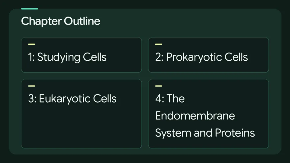
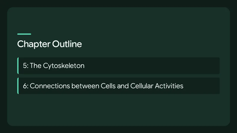
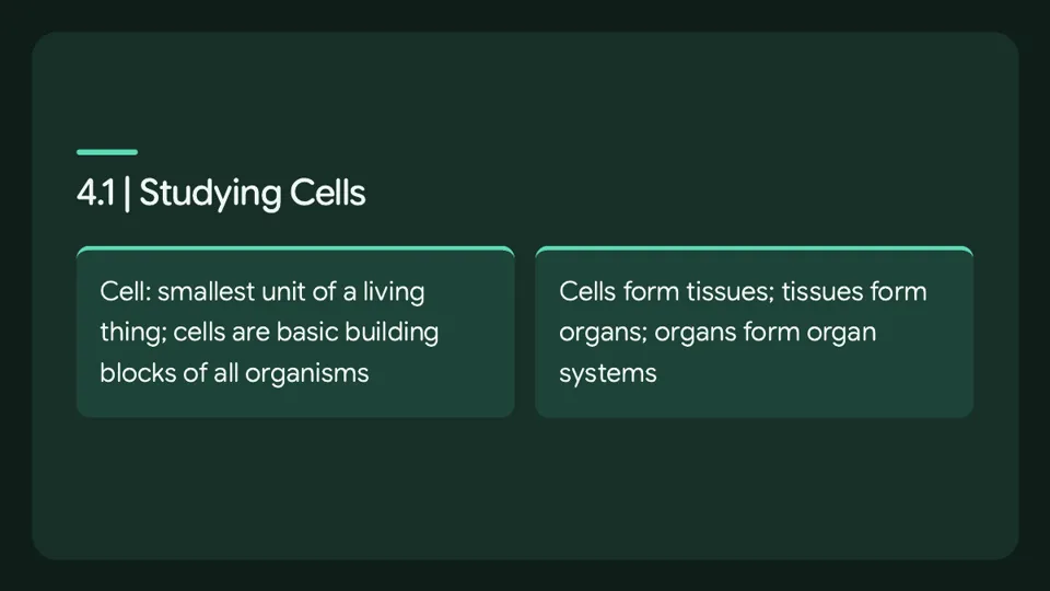
The first slides of lesson 1

  • Slide 1 of lesson 1
  • Slide 2 of lesson 1
  • Slide 3 of lesson 1

The lessons

YUHINA split the chapter into lessons. Each one has its own summary and quiz, shown below as generated.

Lesson 1: Studying Cells: Microscopy and Cell Theory

The human body, like a brick wall, is built from basic units, and these units are cells.

The body contains many kinds of cells, each specialised for a specific purpose.

A cell is defined as the smallest unit of a living thing, and an organism is a living thing whether it consists of one cell or many cells.

Several cells of one kind that interconnect and perform a shared function form tissues.

A microscope is an instrument that magnifies an object, and micrographs are photographs of cells taken with a microscope.

A specimen that is right-side up and facing right on the microscope slide appears upside-down and facing left when viewed through a microscope, and vice versa.

Light microscopes are advantageous for viewing living organisms, but because individual cells are generally transparent, their components are not distinguishable unless they are coloured with special stains, and staining usually kills the cells.

Magnification is the process of enlarging an object in appearance, whereas resolving power is the ability of a microscope to distinguish two adjacent structures as separate, with higher resolution giving better clarity and detail.

To gain a better understanding of cellular structure and function, scientists typically use electron microscopes, although the method used to prepare the specimen for viewing with an electron microscope kills the specimen.

Electrons have short wavelengths, shorter than photons, that move best in a vacuum, so living cells cannot be viewed with an electron microscope.

In a scanning electron microscope, a beam of electrons moves back and forth across a cell’s surface, creating details of cell surface characteristics, while in a transmission electron microscope, the electron beam penetrates the cell and provides details of a cell’s internal structures.

In the 1670s, van Leeuwenhoek discovered bacteria and protozoa.

By the late 1830s, botanist Matthias Schleiden and zoologist Theodor Schwann were studying tissues and proposed the unified cell theory, which states that all living things are composed of one or more cells, the cell is the basic unit of life, and new cells arise from existing cells.

Cytotechnologists are professionals who study cells via microscopic examinations and other laboratory tests, and when abnormalities are discovered early, a patient’s treatment can begin sooner, which usually increases the chances of a successful outcome.

4.1 | Studying Cells

A cell is the smallest unit of a living thing, and a living thing, whether made of one cell like bacteria or many cells like a human, is called an organism, so cells are the basic building blocks of all organisms.

Several cells of one kind that interconnect and perform a shared function form tissues, several tissues combine to form an organ such as the stomach, heart, or brain, and several organs make up an organ system such as the digestive system, circulatory system, or nervous system.

There are many types of cells, all grouped into one of two broad categories, prokaryotic and eukaryotic, and both animal and plant cells are classified as eukaryotic cells, whereas bacterial cells are classified as prokaryotic.

With few exceptions, individual cells cannot be seen with the naked eye, so scientists use microscopes to study them.

Microscopy

A microscope is an instrument that magnifies an object, and most photographs of cells are taken with a microscope, and these images can also be called micrographs.

Light Microscopes — selected points

In light microscopes, visible light passes and is bent through the lens system to enable the user to see the specimen.

Light microscopes commonly used in the undergraduate college laboratory magnify up to approximately 400 times, and magnification is the process of enlarging an object in appearance.

When oil immersion lenses are used for the study of small objects, magnification is usually increased to 1,000 times.

Electron Microscopes

In contrast to light microscopes, electron microscopes use a beam of electrons instead of a beam of light, which allows for higher magnification and thus more detail and also provides higher resolving power.

Cell Theory

In a 1665 publication called Micrographia, experimental scientist Robert Hooke coined the term “cell” for the box-like structures he observed when viewing cork tissue through a lens.

Despite the limitations of his now-ancient lenses, van Leeuwenhoek observed the movements of protista, a type of single-celled organism, and sperm, which he collectively termed “animalcules.”

By the late 1830s, botanist Matthias Schleiden and zoologist Theodor Schwann proposed the unified cell theory, and Rudolf Virchow later made important contributions to this theory.

Cytotechnologist

In this test, a doctor takes a small sample of cells from the uterine cervix of a patient and sends it to a medical lab where a cytotechnologist stains the cells and examines them for any changes that could indicate cervical cancer or a microbial infection.

Cytotechnologists are professionals who study cells via microscopic examinations and other laboratory tests, and they are trained to determine which cellular changes are within normal limits and which are abnormal.

When cytotechnologists notice abnormalities, they consult a pathologist, who is a medical doctor who can make a clinical diagnosis.

4.2 | Prokaryotic Cells

Only the predominantly single-celled organisms of the domains Bacteria and Archaea are classified as prokaryotes.

Cells of animals, plants, fungi, and protists are all eukaryotes and are made up of eukaryotic cells.

Quiz (7 questions)

  1. Which term is defined as “the smallest unit of a living thing”?

    • Cell
    • Cytotechnologists
    • Microscope
    • Tissues
    Show the answer

    Cell. A cell is the smallest unit of a living thing, the basic building block of all organisms. Cytotechnologists are professionals who examine cells, not the smallest unit itself. A microscope is an instrument that magnifies an object, not a unit of life. Tissues are groups of similar cells that work together, which are larger and more complex than a single cell.

  2. “a living thing, whether made of one cell or many cells” — which term does this describe?

    • Magnification
    • Organism
    • Cytotechnologists
    • Cell
    Show the answer

    Organism. An organism is a living thing, whether it is made of one cell or many cells. Magnification is the process of enlarging an object in appearance, not a living thing. Cytotechnologists are professionals who study cells, not the living thing itself. A cell is the smallest unit of a living thing, not the whole living thing made of one or many cells.

  3. Which term is defined as “several systems that function together”?

    • Tissues
    • Cytotechnologists
    • Organism
    • Microscope
    Show the answer

    Organism. Several systems that function together form an organism, such as a human being. Tissues are groups of similar cells that perform a shared function, not a collection of systems. Cytotechnologists are professionals who study cells, not a level of organization. A microscope is an instrument that magnifies an object, not a level of biological organization.

  4. True or false: In a 1665 publication called Micrographia, Robert Hooke coined the term "cell" for the box-like structures he observed when viewing cork tissue through a lens.

    • True
    • False
    Show the answer

    True. Robert Hooke did coin the term "cell" in his 1665 publication Micrographia for the box-like structures he saw in cork tissue. The statement is therefore true, not false.

  5. True or false: The method used to prepare the specimen for viewing with an electron microscope preserves the specimen alive.

    • True
    • False
    Show the answer

    False. The method used to prepare specimens for electron microscopy kills the specimen, so living cells cannot be viewed. The statement is therefore false, not true.

  6. True or false: In a scanning electron microscope, a beam of electrons moves back and forth across a cell's surface, creating details of cell surface characteristics.

    • True
    • False
    Show the answer

    True. In a scanning electron microscope, a beam of electrons moves back and forth across a cell's surface to reveal surface details. The statement is therefore true, not false.

  7. What does “microscope” mean?

    • the smallest unit of a living thing
    • several systems that function together
    • the process of enlarging an object in appearance
    • an instrument that magnifies an object
    Show the answer

    an instrument that magnifies an object. A microscope is an instrument that magnifies an object so that small structures can be seen. "Several systems that function together" describes an organism, not a microscope. "The process of enlarging an object in appearance" is magnification, not the instrument itself. "The smallest unit of a living thing" is a cell, not a microscope.

Lesson 2: Prokaryotic Cells: Structure and Size Constraints

A prokaryote is a simple organism that is mostly single-celled and lacks a nucleus and any other membrane-bound organelle.

Most prokaryotes possess a peptidoglycan cell wall, and many also have a polysaccharide capsule.

The cell wall provides an extra layer of protection, helps the cell maintain its shape, and prevents dehydration, while the capsule enables the cell to attach to surfaces in its environment.

Some prokaryotes have flagella, pili, or fimbriae: flagella are used for locomotion, pili are used to exchange genetic material during a type of reproduction called conjugation, and fimbriae are used by bacteria to attach to a host cell.

Prokaryotic cells measure 0.1 to 5.0 μm in diameter and are significantly smaller than eukaryotic cells, which have diameters ranging from 10 to 100 μm.

The small size of prokaryotes allows ions and organic molecules that enter them to diffuse quickly to other parts of the cell, and wastes produced within a prokaryotic cell can likewise diffuse out quickly.

As a cell increases in size, its surface area-to-volume ratio decreases, so if the cell grows too large the plasma membrane will not have sufficient surface area to support the rate of diffusion required for the increased volume, and the cell becomes less efficient.

One way for a cell to become more efficient is to divide, and another way is to develop organelles that perform specific tasks, since organelles allow different functions to be compartmentalized in different areas of the cell.

4.3 | Eukaryotic Cells — selected points

Unlike prokaryotic cells, eukaryotic cells have a membrane-bound nucleus, numerous membrane-bound organelles such as the endoplasmic reticulum, Golgi apparatus, chloroplasts, mitochondria, and others, and several rod-shaped chromosomes.

Quiz (6 questions)

  1. “a simple, mostly single-celled (unicellular) organism that lacks a nucleus, or any other membrane-bound organelle” — which term does this describe?

    • Microbiologists
    • Organelle
    • Microbes
    • Prokaryote
    Show the answer

    Prokaryote. A prokaryote is a simple, mostly single-celled organism that lacks a nucleus and any other membrane-bound organelle. Microbiologists are scientists who study microbes. An organelle is a small structure inside a cell that performs a specialized job. Microbes are organisms so tiny they can only be seen with microscopes.

  2. “organisms so tiny that they can only be seen with microscopes” — which term does this describe?

    • Microbes
    • Microbiologists
    • Organelle
    • Prokaryote
    Show the answer

    Microbes. Microbes are organisms so tiny that they can only be seen with microscopes, and most are beneficial rather than disease-causing. A prokaryote is a simple, mostly single-celled organism that lacks a nucleus and membrane-bound organelles. Microbiologists are scientists who study microbes. An organelle is a small structure inside a cell that performs a specialized job.

  3. “scientists who study microbes” — which term does this describe?

    • Microbes
    • Microbiologists
    • Prokaryote
    • Organelle
    Show the answer

    Microbiologists. Microbiologists are scientists who study microbes. Microbes are the tiny organisms themselves, which can only be seen with microscopes. A prokaryote is a simple, mostly single-celled organism that lacks a nucleus and membrane-bound organelles. An organelle is a small structure inside a cell that performs a specialized job.

  4. True or false: Prokaryotic cells are 0.1 to 5.0 μm in diameter, significantly smaller than eukaryotic cells, which have diameters ranging from 10 to 100 μm.

    • True
    • False
    Show the answer

    True. Prokaryotic cells measure 0.1 to 5.0 μm in diameter, while eukaryotic cells range from 10 to 100 μm, so prokaryotes are significantly smaller. This small size lets ions, organic molecules, and wastes quickly diffuse, unlike eukaryotic cells, which have structural adaptations to enhance intracellular transport.

  5. True or false: Most prokaryotes have a peptidoglycan cell wall and many have a polysaccharide capsule.

    • True
    • False
    Show the answer

    True. Most prokaryotes have a peptidoglycan cell wall, and many have a polysaccharide capsule. The wall protects the cell, maintains its shape, and prevents dehydration, while the capsule enables attachment to surfaces. The wall and the capsule are distinct structures with different roles.

  6. True or false: Pili are used to exchange genetic material during a type of reproduction called conjugation.

    • True
    • False
    Show the answer

    True. Pili are used to exchange genetic material during a type of reproduction called conjugation. Flagella are used for locomotion, and fimbriae are used by bacteria to attach to a host cell. Only some prokaryotes have these structures.

Lesson 3: Eukaryotic Cell Overview: Plasma Membrane, Cytoplasm, and Nucleus

The plasma membrane regulates what enters and exits the cell, governing the movement of organic molecules, ions, water, and oxygen, and it also allows wastes such as carbon dioxide and ammonia to leave the cell.

The cytoplasm contains dissolved ions of sodium, potassium, calcium, and many other elements, and many metabolic reactions, including protein synthesis, occur there.

During the growth and maintenance phases of the cell cycle, proteins attach to chromosomes, forming unwound, jumbled thread-like complexes known as chromatin, a term that describes the material of chromosomes in both condensed and decondensed states.

Because protein synthesis is essential for all cells, producing enzymes, hormones, antibodies, pigments, structural components, and surface receptors, ribosomes are present in practically every cell.

The Plasma Membrane — selected points

Cells specialized for absorption have plasma membranes folded into fingerlike projections called microvilli, and such cells typically line the small intestine, the organ that absorbs nutrients from digested food.

In celiac disease, an immune response to gluten, a protein in wheat, barley, and rye, damages the microvilli, so afflicted individuals cannot absorb nutrients, which leads to malnutrition, cramping, and diarrhea.

Patients with celiac disease must follow a gluten-free diet.

The Cytoplasm — selected points

The cytoplasm is made up of organelles suspended in the gel-like cytosol, the cytoskeleton, and various chemicals.

Although the cytoplasm is 70 to 80 percent water, proteins within it give it a semi-solid consistency, and the cytoplasm also contains other organic molecules, including glucose and other simple sugars, polysaccharides, amino acids, nucleic acids, fatty acids, and derivatives of glycerol.

The Nucleus

The nucleus is typically the most prominent organelle in a cell.

The nucleus houses the cell's DNA and directs the synthesis of ribosomes and proteins.

The Nuclear Envelope

The nuclear envelope is a double-membrane structure forming the outermost portion of the nucleus, and both its inner and outer membranes are phospholipid bilayers.

Pores punctuate the nuclear envelope and control the passage of ions, molecules, and RNA between the nucleoplasm and the cytoplasm.

The nucleoplasm is the semi-solid fluid inside the nucleus, where the chromatin and the nucleolus are found.

Chromatin and Chromosomes

Chromosomes are structures within the nucleus made up of DNA, the hereditary material.

In prokaryotes, DNA is organized into a single circular chromosome, whereas in eukaryotes chromosomes are linear structures.

Every eukaryotic species has a specific number of chromosomes in the nuclei of its body's cells; for example, humans have 46 and fruit flies have eight.

Chromosomes are visible and distinguishable from one another only when the cell is getting ready to divide.

During the growth and maintenance phases of its life cycle, a cell has proteins attached to its chromosomes, which resemble an unwound, jumbled bunch of threads.

These unwound protein-chromosome complexes are called chromatin ( Figure 4.12); chromatin describes the material that makes up the chromosomes both when condensed and decondensed.

The Nucleolus

Some chromosomes have sections of DNA that encode ribosomal RNA.

A darkly staining area within the nucleus called the nucleolus aggregates the ribosomal RNA with associated proteins to assemble the ribosomal subunits, which are then transported out through the pores in the nuclear envelope to the cytoplasm.

Ribosomes

Under an electron microscope, ribosomes appear either as clusters called polyribosomes or as single, tiny dots floating freely in the cytoplasm.

Ribosomes may be attached to the cytoplasmic side of the plasma membrane, the cytoplasmic side of the endoplasmic reticulum, or the outer membrane of the nuclear envelope.

Electron microscopy has shown that ribosomes, which are large complexes of protein and RNA, consist of two subunits called large and small.

Ribosomes receive their orders for protein synthesis from the nucleus, where DNA is transcribed into messenger RNA, and the mRNA travels to the ribosomes, which translate the code provided by the sequence of nitrogenous bases in the mRNA into a specific order of amino acids in a protein.

Amino acids are the building blocks of proteins.

Ribosomes are particularly abundant in cells that synthesize large amounts of protein; for example, the pancreas is responsible for creating several digestive enzymes, and the cells that produce these enzymes contain many ribosomes.

Mitochondria

Mitochondria, whose singular form is mitochondrion, are often called the "powerhouses" or "energy factories" of a cell because they are responsible for making adenosine triphosphate (ATP), the cell's main energy-carrying molecule.

ATP represents the short-term stored energy of the cell.

Cellular respiration is the process of making ATP using the chemical energy found in glucose and other nutrients.

In mitochondria, this process uses oxygen and produces carbon dioxide as a waste product.

In fact, the carbon dioxide that you exhale with every breath comes from the cellular reactions that produce carbon dioxide as a byproduct.

In keeping with the theme of form following function, it is important to point out that muscle cells have a very high concentration of mitochondria that produce ATP.

When your cells don't get enough oxygen, they do not make a lot of ATP.

Instead, the small amount of ATP they make in the absence of oxygen is accompanied by the production of lactic acid.

Quiz (7 questions)

  1. Which term matches this description: “a phospholipid bilayer with embedded proteins that separates the internal contents of the cell from its surrounding environment”?

    • Ribosomes
    • Cytoplasm
    • Plasma membrane
    • Mitochondria
    Show the answer

    Plasma membrane. The plasma membrane is a phospholipid bilayer with embedded proteins that separates the cell's internal contents from its surroundings and controls the passage of substances into and out of the cell. Ribosomes are the structures responsible for protein synthesis, not a membrane surrounding the cell. The cytoplasm is the entire region between the plasma membrane and the nuclear envelope, not the boundary itself. Mitochondria are organelles that make ATP, not the outer boundary of the cell.

  2. “the entire region of a cell between the plasma membrane and the nuclear envelope” — which term does this describe?

    • Cellular respiration
    • Plasma membrane
    • Cytoplasm
    • Ribosomes
    Show the answer

    Cytoplasm. The cytoplasm is the entire region of a cell between the plasma membrane and the nuclear envelope, made up of organelles suspended in cytosol along with the cytoskeleton and various chemicals. The plasma membrane is the phospholipid bilayer that forms the cell's outer boundary, not the region inside it. Cellular respiration is the process of making ATP using energy from glucose and other nutrients, not a region of the cell. Ribosomes are the structures that carry out protein synthesis, not the space between the membrane and the nucleus.

  3. Which term matches this description: “the organelle that houses the cell's DNA and directs the synthesis of ribosomes and proteins”?

    • Chromosomes
    • Nucleus
    • Nucleolus
    • Ribosomes
    Show the answer

    Nucleus. The nucleus houses the cell's DNA and directs the synthesis of ribosomes and proteins, and it is typically the most prominent organelle in a cell. Chromosomes are the DNA-containing structures within the nucleus, not the organelle that houses them and directs synthesis. Ribosomes are the structures that actually carry out protein synthesis, not the organelle that directs it. The nucleolus is a darkly staining area within the nucleus that assembles ribosomal subunits, not the whole organelle.

  4. True or false: The mRNA travels to the ribosomes, which translate the code provided by the sequence of the nitrogenous bases in the mRNA into a random order of amino acids in a protein.

    • True
    • False
    Show the answer

    False. The mRNA carries the code from the sequence of nitrogenous bases to the ribosomes, which translate it into a specific order of amino acids in a protein. The order is specific, determined by the mRNA sequence, not random, so the statement is false.

  5. True or false: In eukaryotes, chromosomes are linear structures.

    • True
    • False
    Show the answer

    True. In eukaryotes, chromosomes are linear structures, while in prokaryotes DNA is organized into a single circular chromosome. The statement is therefore true.

  6. True or false: Ribosomes may be attached to the cytoplasmic side of the plasma membrane or the cytoplasmic side of the endoplasmic reticulum and the outer membrane of the nuclear envelope.

    • True
    • False
    Show the answer

    True. Ribosomes may appear as clusters or single dots and can be attached to the cytoplasmic side of the plasma membrane, the cytoplasmic side of the endoplasmic reticulum, and the outer membrane of the nuclear envelope. The statement is therefore true.

  7. Which description fits “nuclear envelope”?

    • the process of making ATP using the chemical energy found in glucose and other nutrients
    • the cellular structures responsible for protein synthesis
    • the entire region of a cell between the plasma membrane and the nuclear envelope
    • a double-membrane structure that constitutes the outermost portion of the nucleus
    Show the answer

    a double-membrane structure that constitutes the outermost portion of the nucleus. The nuclear envelope is a double-membrane structure that forms the outermost portion of the nucleus, with pores that control passage between the nucleoplasm and cytoplasm. The process of making ATP using energy from glucose and other nutrients describes cellular respiration, not the nuclear envelope. The entire region between the plasma membrane and the nuclear envelope describes the cytoplasm, not the nuclear envelope. The cellular structures responsible for protein synthesis are ribosomes, not the nuclear envelope.

Lesson 4: Energy and Metabolic Organelles: Mitochondria, Peroxisomes, and Vesicles

The Centrosome

The centrosome is a microtubule-organizing center located near the nuclei of animal cells.

The centrosome contains a pair of centrioles, which are two structures positioned perpendicular to each other.

Each centriole takes the form of a cylinder made of nine triplets of microtubules.

The centrosome, the organelle in which all microtubules originate, replicates itself before a cell divides, and the centrioles appear to play some role in pulling the duplicated chromosomes to opposite ends of the dividing cell.

The exact function of the centrioles in cell division is not clear, however, because cells whose centrosome has been removed can still divide, and plant cells, which lack centrosomes, are capable of cell division.

Lysosomes

Animal cells possess another set of organelles that plant cells do not have, namely lysosomes.

Lysosomes act as the cell's garbage disposal, whereas in plant cells the digestive processes take place in vacuoles.

Enzymes inside the lysosomes help break down proteins, polysaccharides, lipids, nucleic acids, and even worn-out organelles.

Many reactions that occur in the cytoplasm could not take place at a low pH, which again makes apparent the advantage of compartmentalizing the eukaryotic cell into organelles.

These enzymes are active at a much lower pH than that of the cytoplasm.

Therefore, the pH within lysosomes is more acidic than the pH of the cytoplasm.

The Cell Wall

The cell wall is a rigid covering that protects the cell, provides structural support, and gives the cell its shape, and fungal and protistan cells also have cell walls.

The chief component of prokaryotic cell walls is peptidoglycan, while the major organic molecule in the plant cell wall is cellulose, a polysaccharide made up of glucose units.

Chloroplasts

Like the mitochondria, chloroplasts have their own DNA and ribosomes, but chloroplasts carry out an entirely different function.

Chloroplasts are plant cell organelles that carry out photosynthesis, the series of reactions that use carbon dioxide, water, and light energy to make glucose and oxygen.

Like mitochondria, chloroplasts have outer and inner membranes, but within the space enclosed by a chloroplast's inner membrane lies a set of interconnected and stacked fluid-filled membrane sacs called thylakoids.

Each stack of thylakoids is called a granum, and the fluid enclosed by the inner membrane that surrounds the grana is called the stroma.

Chloroplasts contain a green pigment called chlorophyll, which captures the light energy that drives the reactions of photosynthesis.

Like plant cells, photosynthetic protists also have chloroplasts, and some bacteria perform photosynthesis, but their chlorophyll is not relegated to an organelle.

This is a major difference between plants and animals; plants (autotrophs) are able to make their own food, like sugars, while animals (heterotrophs) must ingest their food.

Endosymbiosis

Symbiosis is a relationship in which organisms from two separate species depend on each other for their survival, and endosymbiosis is a mutually beneficial relationship in which one organism lives inside the other.

Endosymbiotic relationships abound in nature, and microbes that produce vitamin K live inside the human gut.

Scientists have long noticed that bacteria, mitochondria, and chloroplasts are similar in size, and bacteria have DNA and ribosomes, just as mitochondria and chloroplasts do.

Scientists believe that host cells and bacteria formed an endosymbiotic relationship when the host cells ingested both aerobic and autotrophic bacteria (cyanobacteria) but did not destroy them.

Through many millions of years of evolution, these ingested bacteria became more specialized in their functions, with the aerobic bacteria becoming mitochondria and the autotrophic bacteria becoming chloroplasts.

This relationship is beneficial for us because we are unable to synthesize vitamin K.

It is also beneficial for the microbes because they are protected from other organisms and from drying out, and they receive abundant food from the environment of the large intestine.

The Central Vacuole

The central vacuole plays a key role in regulating the cell's concentration of water in changing environmental conditions.

When a plant is left unwatered for a few days, it wilts because, as the water concentration in the soil becomes lower than the water concentration in the plant, water moves out of the central vacuoles and cytoplasm.

As the central vacuole shrinks, it leaves the cell wall unsupported, and this loss of support to the cell walls of plant cells results in the wilted appearance of the plant.

Quiz (7 questions)

  1. Which term is defined as “small, round organelles enclosed by single membranes”?

    • Peroxisomes
    • Glyoxysomes
    • Cell wall
    • Symbiosis
    Show the answer

    Peroxisomes. Peroxisomes are small, round organelles enclosed by single membranes, which matches the definition exactly. The cell wall is a rigid covering outside the plasma membrane that protects and supports the cell, not a small round organelle. Glyoxysomes are specialized peroxisomes in plants that convert stored fats into sugars, not the general term defined here. Symbiosis is a relationship between organisms of two separate species, not an organelle.

  2. Which term is defined as “a microtubule-organizing center found near the nuclei of animal cells”?

    • Peroxisomes
    • Endosymbiosis
    • Glyoxysomes
    • Centrosome
    Show the answer

    Centrosome. The centrosome is a microtubule-organizing center found near the nuclei of animal cells, which is the definition given. Endosymbiosis is a mutually beneficial relationship in which one organism lives inside another, not a cellular structure. Glyoxysomes are specialized peroxisomes in plants that convert stored fats into sugars, not a microtubule-organizing center. Peroxisomes are small, round organelles enclosed by single membranes that carry out oxidation reactions, not a microtubule-organizing center.

  3. “a polysaccharide made up of glucose units” — which term does this describe?

    • Stroma
    • Cellulose
    • Centrosome
    • Symbiosis
    Show the answer

    Cellulose. Cellulose is a polysaccharide made up of glucose units, which is the description given. The stroma is the fluid enclosed by the inner membrane of a chloroplast that surrounds the grana, not a polysaccharide. The centrosome is a microtubule-organizing center found near the nuclei of animal cells, not a polysaccharide. Symbiosis is a relationship in which organisms from two separate species depend on each other, not a polysaccharide.

  4. True or false: The chief component of prokaryotic cell walls is peptidoglycan, while the major organic molecule in the plant cell wall is cellulose.

    • True
    • False
    Show the answer

    True. The chief component of prokaryotic cell walls is peptidoglycan, while the major organic molecule in the plant cell wall is cellulose, so the statement is true. Cellulose is a polysaccharide made up of glucose units, and it is the major organic molecule in the plant cell wall, not in prokaryotic cell walls.

  5. True or false: The pH within lysosomes is more acidic than the pH of the cytoplasm.

    • True
    • False
    Show the answer

    True. The enzymes within lysosomes are active at a much lower pH than that of the cytoplasm, so the pH within lysosomes is more acidic than the pH of the cytoplasm, making the statement true. Lysosomes are the cell's garbage disposal, and their acidic interior is what allows their enzymes to function.

  6. True or false: When confined to peroxisomes, enzymes safely break down the H2O2 released by oxidation reactions into oxygen and water.

    • True
    • False
    Show the answer

    True. Oxidation reactions in peroxisomes release hydrogen peroxide, which would be damaging to cells, but when these reactions are confined to peroxisomes, enzymes safely break down the H2O2 into oxygen and water, so the statement is true. Peroxisomes are small, round organelles enclosed by single membranes that carry out these oxidation reactions.

  7. What does “Chloroplasts” mean?

    • plant cell organelles that carry out photosynthesis
    • specialized peroxisomes in plants responsible for converting stored fats into sugars
    • membrane-bound sacs that function in storage and transport
    • the fluid enclosed by the inner membrane that surrounds the grana
    Show the answer

    plant cell organelles that carry out photosynthesis. Chloroplasts are plant cell organelles that carry out photosynthesis, the series of reactions that use carbon dioxide, water, and light energy to make glucose and oxygen. The fluid enclosed by the inner membrane that surrounds the grana is the stroma, not the chloroplast itself. Specialized peroxisomes in plants responsible for converting stored fats into sugars are glyoxysomes, not chloroplasts. Membrane-bound sacs that function in storage and transport are vesicles and vacuoles, not chloroplasts.

Flashcards

  • cell

    the smallest unit of a living thing

  • organism

    a living thing, whether made of one cell or many cells

  • tissues

    several cells of one kind that interconnect with each other and perform a shared function

  • microscope

    an instrument that magnifies an object

  • micrographs

    photographs of cells taken with a microscope

  • magnification

    the process of enlarging an object in appearance

  • unified cell theory

    all living things are composed of one or more cells, the cell is the basic unit of life, and new cells arise from existing cells

  • cytotechnologists

    professionals who study cells via microscopic examinations and other laboratory tests

  • plasma membrane

    an outer covering that separates the cell's interior from its surrounding environment

  • prokaryote

    a simple, mostly single-celled (unicellular) organism that lacks a nucleus, or any other membrane-bound organelle

  • microbes

    organisms so tiny that they can only be seen with microscopes

  • microbiologists

    scientists who study microbes

  • organelle

    The word “organelle” means “little organ,”

  • cytoplasm

    the entire region of a cell between the plasma membrane and the nuclear envelope

  • nucleus

    the organelle that houses the cell's DNA and directs the synthesis of ribosomes and proteins

  • nuclear envelope

    a double-membrane structure that constitutes the outermost portion of the nucleus

  • chromosomes

    structures within the nucleus that are made up of DNA, the hereditary material

  • nucleolus

    a darkly staining area within the nucleus that aggregates the ribosomal RNA with associated proteins to assemble the ribosomal subunits

  • ribosomes

    the cellular structures responsible for protein synthesis

  • mitochondria

    organelles often called the powerhouses or energy factories of a cell because they are responsible for making adenosine triphosphate (ATP), the cell's main energy-carrying molecule

  • cellular respiration

    the process of making ATP using the chemical energy found in glucose and other nutrients

  • Peroxisomes

    small, round organelles enclosed by single membranes

  • Glyoxysomes

    specialized peroxisomes in plants responsible for converting stored fats into sugars

  • Vesicles and vacuoles

    membrane-bound sacs that function in storage and transport

  • Centrosome

    a microtubule-organizing center found near the nuclei of animal cells

  • Cell wall

    a rigid covering that protects the cell, provides structural support, and gives shape to the cell

  • Cellulose

    a polysaccharide made up of glucose units

  • Chloroplasts

    plant cell organelles that carry out photosynthesis

  • Photosynthesis

    the series of reactions that use carbon dioxide, water, and light energy to make glucose and oxygen

  • Stroma

    the fluid enclosed by the inner membrane that surrounds the grana

  • Symbiosis

    a relationship in which organisms from two separate species depend on each other for their survival

  • Endosymbiosis

    a mutually beneficial relationship in which one organism lives inside the other

Mind map

4.1: Studying Cells 4.2: Prokaryotic Cells 4.3: Eukaryotic Cells

  • Studying Cells

    • Microscopy
      • Light Microscopes
      • Electron Microscopes
    • Cell Theory
      • Unified cell theory
      • Rudolf Virchow
  • Prokaryotic Cells

    • Components of Prokaryotic Cells
      • plasma membrane
      • cytoplasm
      • DNA
      • ribosomes
        • polyribosomes
        • large and small
      • peptidoglycan cell wall
      • polysaccharide capsule
      • flagella
      • pili
      • fimbriae
    • Cell Size
      • surface area-to-volume ratio
  • Eukaryotic Cells

    • The Plasma Membrane
      • phospholipid bilayer
      • microvilli
    • The Cytoplasm
      • cytosol
      • cytoskeleton
    • The Nucleus
    • Animal Cells versus Plant Cells
      • The Centrosome
        • centrioles
      • Lysosomes
      • The Cell Wall
        • cellulose
      • Chloroplasts
        • thylakoids
        • granum
        • stroma

The podcast

The first 90 seconds of a 37-minute episode about the whole chapter. How it opens:
host
Welcome to the episode. I am glad you are joining us. Today we begin a guided tour of the cell, starting with how scientists study cells and what a cell actually is.
guest
Thank you for having me. It is a pleasure to start at the foundation. Before we look at any particular kind of cell, it helps to be clear about what a cell is and how cells fit into the larger organization of a living thing.
host
Then let us start right there. What exactly is a cell?
guest
A cell is the smallest unit of a living thing. A living thing, whether it is made of one cell, like bacteria, or many cells, like a human, is called an organism. In that sense, cells are the basic building blocks of all organisms.
host
And how do those building blocks come together to make something as complex as an organ?
guest
Several cells of one kind that interconnect with each other and perform a shared function form tissues. Several tissues combine to form an organ, such as your stomach, heart, or brain. Several organs then make up an organ system, such as the digestive system, the circulatory system, or the nervous system.

Source: sections 4.1 to 4.3 of chapter 4, “Cell Structure”, from Biology (1st edition) by Connie Rye, Robert Wise et al., OpenStax, licensed CC BY 4.0. Biology. Access the book for free at openstax.org. Changes: YUHINA turned the text into lessons, slides, narration, questions, flashcards, a mind map and a podcast.

Try it with your own chapterSee pricing

More samples

  • Psychology

    Memory

    3 lessons · 57 slides · 124 questions · 26 flashcards · 25-minute podcast

  • Chemistry

    Atoms, Molecules, and Ions

    4 lessons · 54 slides · 112 questions · 17 flashcards · 37-minute podcast

  • Anatomy and Physiology

    Heart Anatomy

    4 lessons · 60 slides · 110 questions · 32 flashcards · 43-minute podcast

  • Astronomy

    The Sun

    4 lessons · 58 slides · 112 questions · 22 flashcards · 43-minute podcast

  • Physics

    Forces and Newton’s Laws of Motion

    4 lessons · 63 slides · 112 questions · 18 flashcards · 44-minute podcast

  • American Government

    The Constitution and Its Origins

    3 lessons · 82 slides · 104 questions · 15 flashcards · 55-minute podcast

  • Sociology

    Culture

    2 lessons · 86 slides · 89 questions · 20 flashcards · 50-minute podcast

  • Economics

    Demand and Supply

    4 lessons · 50 slides · 114 questions · 22 flashcards · 36-minute podcast

All samples
YUHINA

One chapter in. A whole course out.

YUHINA generates the teaching content around your source material — narrated video, question banks, slide decks, podcasts, mind maps and flash cards, all from the same text.

Questions?

Support
support@yuhina.ai
General inquiries
hello@yuhina.ai
Enterprise
enterprise@yuhina.ai

Explore

  • Home
  • Process
  • Outputs
  • Pricing
  • Compare
  • Samples
  • Documentation
  • Contact
  • Get started
  • Sign in
  • Enterprise
  • YouTube
  • X

What it generates

  • PDF to video
  • PDF to quiz
  • PDF to flash cards
  • PDF to podcast
  • PDF to mind map
  • PDF summary

Who it's for

  • Teachers
  • Students
  • Trainers
  • Tutoring centres

Legal

  • Terms
  • Privacy
  • Refunds
  • AI disclosure
  • Accessibility
  • Subprocessors
  • Security
  • This site's privacy notice

© 2026 YUHINA. All rights reserved.

Back to top

YUHINA